New Vision Fundus is the best choice for professional Medical Imaging.
Europe's #1 top selling, FDA 510(k) cleared ophthalmic medical image processing and digital storage software!
Download!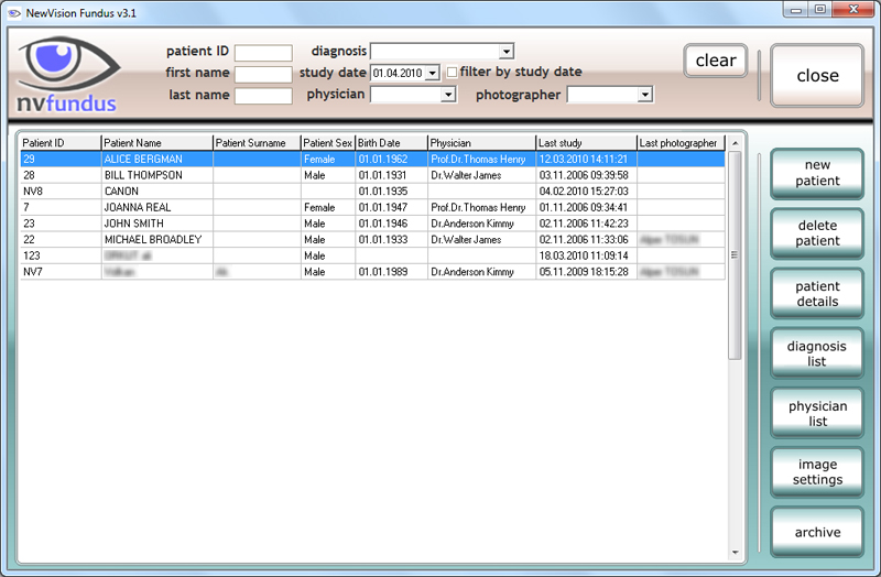
Europe's #1 top selling, FDA 510(k) cleared ophthalmic medical image processing and digital storage software!
Looking for a replacement for Topcon IMAGEnet or Harmony? NewVision Fundus provides a modern, vendor-neutral alternative.
NewVision Fundus is FDA 510(k) cleared for the United States market.
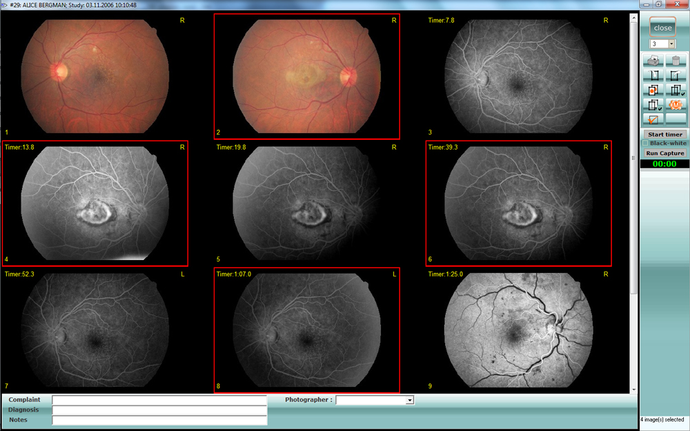
Fundus Fluorescein Angiography (FFA) and ICG angiography are made simple.
NewVision Fundus supports photodynamic therapy (PDT) analysis, planning, treatment, measurement, and documentation.
Various image processing functions provide tools for diverse imaging tasks such as visualization, enhancement, measurement, and documentation.
NewVision is a modular ophthalmic PACS software designed to replace legacy systems like Topcon IMAGEnet and Harmony, enabling seamless integration with fundus cameras and DICOM workflows.
NewVision integrates digital cameras and software and provides convenient means for image acquisition, storage, manipulation, and analysis, as well as data exchange and data backup.
Upgrade your legacy fundus camera without replacing hardware
Vendor-neutral replacement for legacy ophthalmic software
Modernize your workflow, keep your camera investment, and move to a more flexible, vendor-neutral ophthalmic imaging platform.
Topcon NW400, NW500, Topcon TRC series
Canon CR series, Megavision cameras, Allied Vision cameras
Zeiss systems
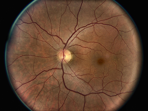
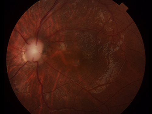
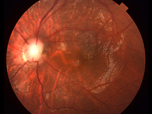
Enhancement Technology in Fundus Images The retinal fundus image plays an important role in the diagnosis of retinal diseases. Detailed information of the retinal fundus image, such as small vessels, microaneurysms, and exudates, may be of low contrast, and retinal image enhancement often helps to analyze retinal fundus image-related diseases. Image enhancement can be applied to any fundus image taken thanks to the "Enhancement" function (image enhancement function) in New Vision Fundus. A new image can be saved by applying the desired image enhancement process to the images transferred to the New Vision Fundus. The “Enhancement” functions help the doctor to examine the fundus images taken with the ophthalmic camera in more detail. According to the feedback we received from our customers, it seems that the “Enhancement” functions benefit both doctors and patients. Thanks to the image enhancement process, images that are too dark to be evaluated are improved and brought to a level that can be analyzed by the doctor.

Auto Contrast Button
Automatic Enhancement Button


Haze Enhancement Button

Cataract FA Enhancement Button


Color Enhancement Button


Automatic Enhancement Button

Automatic Enhancement (Medium) Button


Automatic Enhancement (Aggressive)
CE APPROVED OPHTHALMIC MEDICAL IMAGE PROCESSING AND DIGITAL STORAGE SOFTWARE
Overview New Vision DICOM Server is a complete DICOM Solution enabling seamless integration of all types of DICOM-enabled automated microscopes and digital cameras with New Vision Medical Imaging Software, leading ultimately to a complete Hospital PACS system. New Vision DICOM Server offers full-featured DICOM support and functionality.
NewVision Fundus DICOM Server is a Level 2 (Full) Service Class Provider:
Sales Office, San Francisco - Galvanize, Innogate Suite 409, 44 Tehama St, San Francisco, CA 94105, United States
R&D Center, Turkey - Pınarbaşı Mah. Hürriyet Cad. Akdeniz Üniversitesi Antalya Teknokent Ar-Ge 2 Uluğbey Apt, No: 3A / Z14 Konyaaltı / ANTALYA / TÜRKİYE
info @ uraltelekom.com
Fundus Photo - The exclusive dealer for North America, South America and Central America.